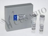
Phospho-Histone H3.3 (T3) AntibodyСпецификация| Объем | 100 мкл | | Синонимы | Histone H3.3, H3F3A, H3.3A, H3F3, PP781, AND, H3F3B, H3.3B | | Тип антител | Recombinant Antibody | | Species | Human | | UniProt ID | P84243 | | Иммуноген | A synthesized peptide | | Видовая специфичность | Human ,Mouse | | Применение | ELISA, WB, ICC, FC, Recommended dilution: WB:1:500-1:5000, ICC:1:50-1:500 | | Клональность | Monoclonal | | Изотип | Rabbit IgG | | Коньюгат | Non-conjugated | | Буффер | Rabbit IgG in phosphate buffered saline, pH 7.4, 150mM NaCl, 0.02% sodium azide and 50% glycerol. | | Форма | Liquid | | Хранение | Upon receipt, store at -20°C or -80°C. Avoid repeated freeze. | | Метод очистки | Affinity-chromatography | | Области исследований | Epigenetics and Nuclear Signaling | | Аббревиатура | Histone H3.3 | | Примечание | Variant histone H3 which replaces conventional H3 in a wide range of nucleosomes in active genes. Constitutes the predominant form of histone H3 in non-dividing cells and is incorporated into chromatin independently of DNA synthesis. Deposited at sites of nucleosomal displacement throughout transcribed genes, suggesting that it represents an epigenetic imprint of transcriptionally active chromatin. Nucleosomes wrap and compact DNA into chromatin, limiting DNA accessibility to the cellular machineries which require DNA as a template. Histones thereby play a central role in transcription regulation, DNA repair, DNA replication and chromosomal stability. DNA accessibility is regulated via a complex set of post-translational modifications of histones, also called histone code, and nucleosome remodeling. | | Ссылка на страницу на сайте производителя | ссылка | Western Blot
Positive WB detected in:Hela whole cell lysate,293 whole cell lysate,NIH/3T3 whole cell lysate
All lanes:Phospho-Histone H3 (T3) antibody at 1.41µg/ml
Secondary
Goat polyclonal to rabbit IgG at 1/50000 dilution
Predicted band size: 16 KDa
Observed band size: 16 KDa
 | Immunocytochemistry analysis of CSB-RA010109A03phHU diluted at 1:100 and staining in Hela cells performed on a Leica BondTM system. After dewaxing and hydration, antigen retrieval was mediated by high pressure in a citrate buffer (pH 6.0). Section was blocked with 10% normal goat serum 30min at RT. Then primary antibody (1% BSA) was incubated at 4°C overnight. The primary is detected by a biotinylated secondary antibody and visualized using an HRP conjugated SP system.
 | Overlay histogram showing Hela cells stained with CSB-RA010109A03phHU (red line) at 1:50. The cells were fixed with 70% Ethylalcohol (18h) and then permeabilized with 0.3% Triton X-100 for 2 min.The cells were then incubated in 1x PBS /10% normal goat serum to block non-specific protein-protein interactions followed by primary antibody for 1 h at 4?.The secondary antibody used was FITC goat anti-rabbit IgG (H+L) at 1/200 dilution for 1 h at 4?. Control antibody (green line) was used under the same conditions. Acquisition of >10,000 events was performed.
 | | | |
Информация для заказа| Область использования: | Производство: | Cusabio | | Метод: | Антитела | | Объем: | 100 мкл | | Кат. номер: | CSB-RA010109A03phHU | | Цена (с НДС 20%): | по запросу | В корзину  |  Наименование: Phospho-Histone H3.3 (T3) Antibody. Наименование: Phospho-Histone H3.3 (T3) Antibody.
Примечание: дополнительная информация (на английском языке). |
|
